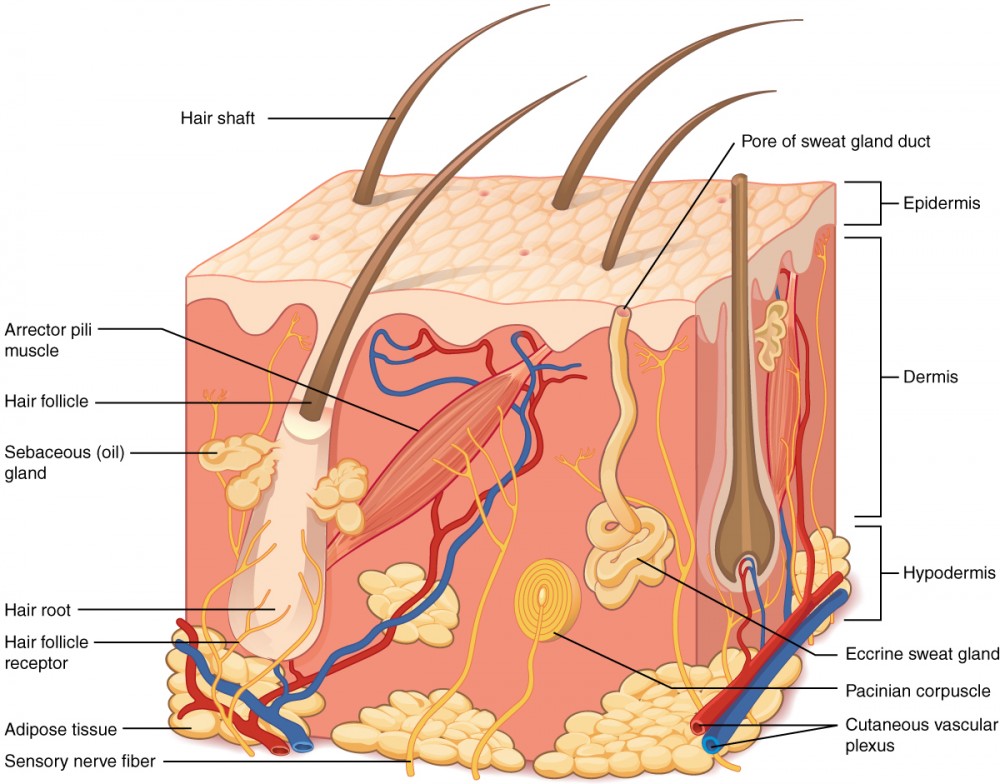
The Nerve Fibers in the Dermis Stimulate
The skin is innervated by afferent somatic nerves with fine unmyelinated c or myelinated aδ primary afferent nerve fibers transmitting sensory stimuli temperature changes chemicals inflammatory mediators ph changes via dorsal root ganglia and the spinal cord to specific areas of the central nervous system cns resulting in the. The ways in which skin promotes loss of excess body heat are _____.
Layers Of The Epidermis Youtube Layers Of The Epidermis Epidermis Skin Anatomy
However at painful intensities both non-nociceptive Aβ-fibers and nociceptive Aδ- and C-fibers may be activated by the electrical stimulation.

. The principal types of fibers are types I and III collagen and elastic fibers. Electrical stimulation of cutaneous tissue through surface electrodes is an often used method for evoking experimental pain. Muscles and glands in the dermis.
Nerve endings in the dermis surround hair follicles. The nerve fibers in the dermis stimulate A. What are the arrector pili muscles responsible for.
You step out of the shower and vigorously rub your skin with a towel. Body heat is lost by radiation by. They are most likely.
The attempt to remove their finger prints. When internal organs need more blood or more heat nerves stimulate the dermal vessels to constrict shunting more blood into the general circulation and. The concentration of substance P SP and the density of SP-immunoreactive nerve fibers in dermis are known to increase during wound healing 66.
75 rows The nerve fibers in the dermis stimulate. Muscles and glands in the dermis. The major blood vessels that supply the skin are in the.
Skin of AD lesion is hyper-innervated with increased SP- and CGRP-positive nerve fibers in the epidermis and papillary dermis with increased mast cell-nerve fiber contacts. The police recover the jewelry and an officer explains on the evening news the fingerprints were obtained from the back of the watch. Muscles and glands in the dermis.
The nerve fibers in the dermis stimulate what. Secrete synovial fluid that lubrictes the ends of bones at joints. Blood vessels in the epidermis.
What motor nerves stimulate the arrector pili muscles. If you were able to analyze the towel you would find skin cells. Cultured papillary FBs are characterized by an increased expression of GM-CSF upon stimulation although expression of this factor is the same for cells of both layers of the dermis in vivo.
This study proposes a. The major blood vessels that supply the. Shortly after the eighth week sensory nerves growing into the dermis and epidermis help complete reflex arcs and thus allow the fetus to respond to.
2 theives rob jewelry store. IES is based on the fact that nociceptive fiber terminals are located in the epidermis whereas receptors of other fibers end deep in the dermis. Nerve fibers in the dermis stimulate Muscles and glands in the dermis Two thieves steal jewelry and then drop it as they are escaping.
Deep pressure receptors also exist. Two thieves steal jewelry and then. Sweat glands blood vessels and the arrector pili muscle are innervated by sympathetic C-fibers in the dermis.
The prints arise form the dermis which is not destroyed. The dermal blood vessels consist of two vascular plexuses a plexus is a network of converging and diverging vessels. Why does this not work.
Due to the concentric geometric shape and short anodecathode distance a high current density can be achieved at relatively low current intensities causing the selective depolarization of nociceptive fibers in the superficial layer of the dermis without recruiting deeper nonnociceptive thick fibers. The skin is innervated by afferent somatic nerves with fine unmyelinated c or myelinated aδ primary afferent nerve fibers transmitting sensory stimuli temperature changes chemicals inflammatory mediators ph changes via dorsal root ganglia and the spinal cord to specific areas of the cns resulting in the perception of pain burning. The dermis becomes highly vascularized with an early capillary network transformed into layers of larger vessels.
Melanocytes in the epidermis. Abstract Intra-epidermal electric stimulation IES is an alternative to laser stimulation for selective activation of cutaneous Aδ-fibers. Fat cells in the subcutaneous layer.
In addition papillary and reticular FBs. Muscles the glands in the dermis. Calcitonin gene-related peptide and SP have been shown to stimulate fibroblasts and neurogenic inflammation as well as epidermal organization and keratinocyte renewal 67.
Afferent intraepidermal nerve fibers of the class C and Aδ are found in the epidermis as free nerve endings. These nerve endings sense hair movement and act as mechanoreceptors allowing sensation to extend beyond the skins surface. Radiation conduction convection evaporation.
The dermis is richly supplied with nerve fiber and blood vessels. In epidermis neuropeptides released from the nerve fibers stimulate keratinocytes to produce proinflammatory cytokines such as IL-1α. The nerve fibers in the dermis function to stimulate _____.
Muscles and glands in the dermis.
Concept Map Integumentary If You Need Help Turning Javascript On Click Here Integumentary System Concept Map Nursing Study Guide
Transduction Of An Itch In The Skin A C A Degenerating Download Scientific Diagram
Biomedicines Free Full Text Design Of In Vitro Hair Follicles For Different Applications In The Treatment Of Alopecia A Review Html
Pin By Carsen Ortiz On Cosmetology Beauty In 2021 Sensory Nerves Dermis Nerve Fiber
Human Skin Anatomy Epidermis With In 2022 Skin Anatomy Epidermis Human
Pgp9 5 Staining Of Nerve Fibers In The Epidermis And Upper Dermis Download Scientific Diagram
Layers Of The Skin Anatomy And Physiology
Click The Link In Our Bio To Learn About 6 Types Of Sensory Receptors Neuroanatomy Neuroscience Histology Medical School Studying Sensory Science Biology
Intra Epidermal Free Nerve Endings The Epidermis Is Innervated By Download Scientific Diagram
A Simplified Illustration Of The General Anatomy Of The Skin With The Download Scientific Diagram
Seer Training Anatomy Of The Skin
Al Shaafi Herbal Hair Oil 50 Oils In A Bottle With 60 Etsy Integumentary System Herbal Hair Oils Skin Anatomy
Three Dimensional Imaging Of The Hyperpigmented Skin Of Senile Lentigo Reveals Underlying Higher Density Intracutaneous Nerve Fibers Journal Of Dermatological Science
Comments
Post a Comment